
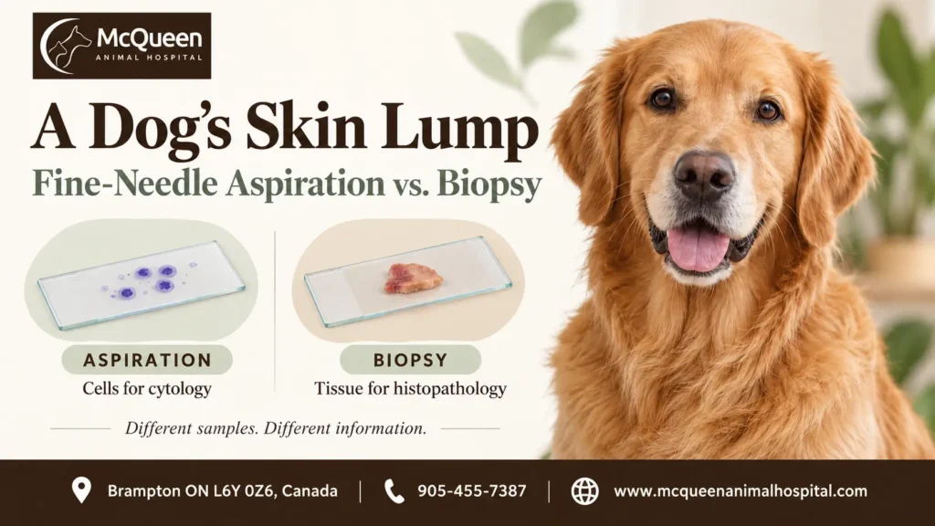
Fine-needle aspiration usually collects individual cells from a dog’s lump for cytology, while a tissue biopsy collects a piece of the lump for histopathology. Cytology examines what the cells look like; histopathology also shows how they are arranged within the tissue. The more useful test depends on the lump and the question your veterinarian needs to answer.
A recommendation to sample a lump does not mean your dog has cancer. Cysts, inflammation and benign growths can also cause lumps in or beneath the skin. Sampling helps distinguish these possibilities and identify when further investigation is needed.
Cells versus tissue: what each test can show
During fine-needle aspiration, sometimes called a dog lump needle test, the veterinarian uses a thin needle to collect material for examination on microscope slides. The sample may contain cells, fluid or other material from the lump. Fine-needle samples can also be collected without suction, although owners may hear both approaches described as a needle aspirate.
Cytology can sometimes identify a particular condition, such as a fatty growth called a lipoma or certain types of tumour. In other cases, it identifies a broad category of cells or inflammation without establishing exactly what the lump is. Its value in classifying skin growths is described in the veterinary pathology review Cytology of Skin Neoplasms.
A tissue biopsy preserves more of the cells’ surroundings. Instead of seeing mainly loose cells, the pathologist can examine their organisation and relationship to neighbouring tissue—known as tissue architecture. Depending on the sample and diagnosis, this may help establish whether a growth is invading surrounding tissue or provide a tumour grade, a microscopic assessment relevant to some tumour types. AAHA’s oncology guidance explains why these details can require histopathology even when cytology has already identified abnormal cells.
| Practical difference | Fine-needle aspiration and cytology | Tissue biopsy and histopathology |
|---|---|---|
| What is collected? | Cells and other material through a thin needle | A piece of tissue, or sometimes the whole lump |
| Main information | Cell appearance; sometimes a specific diagnosis | Cell appearance plus tissue structure |
| What might remain unanswered? | Exact lump type, invasion or grade | Questions requiring a more representative sample or additional laboratory tests |
| Procedure involved | Usually less invasive; some dogs need sedation | Requires an individual pain-control and anaesthesia plan; wound care may be needed |
| What happens next? | Monitoring, further sampling or another decision based on the findings | Further decisions based on the diagnosis and any remaining uncertainty |
The terminology can overlap: “fine-needle biopsy” sometimes refers to cell sampling. If your veterinarian recommends a biopsy, ask whether they mean a needle sample for cytology or a tissue sample for histopathology.
Why your veterinarian might choose one method first
Fine-needle aspiration is often a useful starting point because it can provide information with relatively little disruption to the lump. A clear result may answer the immediate question or help the veterinarian plan the next procedure. However, some lumps release few cells, and some diagnoses depend on tissue structure that a cell sample cannot show.
A tissue biopsy may therefore be recommended first when the veterinarian expects cytology to leave an important question unresolved. It may also follow an aspirate that identifies a growth but does not provide enough detail for the next decision. During the veterinary consultation and examination, the veterinarian considers the lump’s location, size, consistency and changes over time, alongside your dog’s health and any previous results.
A biopsy does not necessarily mean removing the entire lump. An incisional biopsy takes part of it; an excisional biopsy removes the whole visible lump. The veterinarian should explain which approach is proposed and why, including whether establishing the diagnosis beforehand could affect how removal is planned. McQueen lists lumpectomy among its pet surgery services, but whether removal is appropriate depends on the individual lump and assessment.
If you have not yet had the lump examined, arrange a veterinary assessment at an animal hospital and bring any photographs or notes showing when you noticed it and how it has changed.
What the sampling appointment may involve
For a fine-needle aspirate, your dog needs to remain sufficiently still for safe sampling. Some dogs tolerate this while awake, but sedation may be appropriate because of anxiety, discomfort, movement or the lump’s location. Do not assume that a needle test will be painless or that every dog can have it without sedation; ask how your dog’s comfort will be managed.
A tissue biopsy may involve a skin punch, a core of tissue or a surgical incision. Local anaesthesia, sedation or general anaesthesia may be used, depending on the technique, site and patient. If general anaesthesia is proposed, McQueen’s information about pet anaesthesia and monitoring provides context for that discussion. Your dog may need stitches and instructions about activity, licking and wound checks afterwards.
Before either procedure, tell the clinic about medications, supplements, previous anaesthetic problems and any history of unusual bleeding. Ask whether your dog needs to fast and whether usual medicines should be given. Follow the treating clinic’s instructions rather than withholding food or medication on your own.
What an inconclusive result means
A non-diagnostic sample does not contain enough useful material to establish a diagnosis. An inconclusive result may contain interpretable cells or tissue but still leave more than one possible explanation. Neither result establishes that a lump is harmless.
A needle sample may contain too few cells or fail to represent the important part of the lump. In a retrospective study of skin and subcutaneous masses from dogs and cats, some aspirates could not be evaluated because they contained too few cells; other cytology findings differed from the subsequent tissue diagnosis. This illustrates why an aspirate cannot always exclude a tumour.
The next step depends on what prevented an answer. Repeating the aspirate from another part of the lump may help if the first sample was unrepresentative. A tissue biopsy may be more useful if the cells do not release readily or their appearance alone cannot distinguish the possibilities.
Biopsy also has limits. A small piece may not capture every component of a varied lump, and some tissue samples require additional stains or other laboratory tests to classify them. A recommendation for another test should come with an explanation of the unanswered question and how the result could change the plan.
Who reviews the sample—and what happens after the report?
Collecting the sample and interpreting it are separate steps. A veterinarian may examine cytology slides in the clinic, while a diagnostic laboratory can provide review by a veterinary clinical pathologist. Tissue samples undergo laboratory processing for histopathology. Ask whether the result you receive is a preliminary assessment or the completed laboratory report.
McQueen’s in-house laboratory page describes checking samples from abnormal growths, including aspirates and biopsies, before sending them to a pathologist. This sample-quality check is distinct from the pathologist’s final interpretation.
Before proceeding, useful questions include:
- What are we trying to establish with this sample?
- Who will review it, and is laboratory interpretation included in the estimate?
- What preparation and aftercare does my dog need?
- When should I expect an update, and whom should I contact if it has not arrived?
- If the result is unclear, what would you recommend next—and why?
When the report arrives, ask how confidently it identifies the lump and whether it fits the examination findings. If monitoring is recommended, agree on what to measure, when to recheck and which changes should prompt earlier contact. The useful outcome is a clear understanding of what the sample established, what remains uncertain and what decision comes next.
Medically reviewed by Dr. Harinder Babbar, DVM
Veterinarian & Medical Director
McQueen Animal Hospital, Brampton, OntarioThis article has been medically reviewed and is based on current evidence-based veterinary literature and clinical guidance.