On-Site Pet Ultrasonography
veterinary ultrasonography brampton
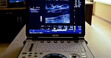
Ultrasound is an invaluable instrument in the detection of abnormalities in our pets. It non-invasively provides a detailed and very accurate look inside the abdomen and heart, detecting even small changes well before other tests such as x-rays or blood tests would. It is important to note that not all ultrasound equipment is considered equal. We invested in equipment from GE, world leaders in human and veterinary ultrasonography.
The list of uses for medical ultrasound is extremely long, however common uses include pregnancy checks, examination of the urinary bladder (for stones, crystals, infection, cancer, and more), examination of the kidneys (for infection, congenital defects, cancer, stones, and more), liver (for various forms of liver disease, cancer, gall bladder disease or stones, and more), and the list goes on and on. Our veterinarian has gone through extensive training to be able to provide this service to you right here in our hospital, the moment your pet needs it.
Organs can be sampled with ultrasound guidance, as illustrated below, allowing the diagnosis and treatment of many organ diseases without invasive procedures such as surgical biopsies.
Case Studies
How pet ultrasound helps
Even in the short period of time that our clinic has been opened, we have already had many patients that we’ve been able to help with the use of our ultrasound, allowing us to get much needed answers for our clients and their beloved pets. Below are a few examples.
Lucky – 2 Year Old Shih-Tzu
Lucky presented to us with lethargy, weight loss, and anorexia. In-house blood tests performed immediately revealed significantly abnormal changes to kidney values. To characterize what type of kidney disease was present, an abdominal ultrasound was recommended. This is because many disease processes can cause changes in blood tests, including cancer, kidney failure, toxins, dehydration and more.
Unfortunately, Lucky was found to have renal dysplasia based on ultrasound findings. Renal dysplasia refers to the improper development of kidneys that is often a genetic abnormality, particularly in this breed. Sadly, there is no cure for this particular disease, but it can be managed with appropriate diet and medications. Ultrasound allowed us to immediately determine a reason for the elevated kidney enzymes on blood tests, and allowed us to avoid further unnecessary and potentially expensive testing.
Lola – 8 Year Old Labrador Retriever

Lola presented to us after being treated by another veterinary hospital. There, Lola had blood tests done which led them to thinking she had an endocrine disease called Cushing’s, where the adrenal glands produce excessive amounts of the stress hormone, cortisol. Because Lola lacked some of the clinical signs we expect for this disease, an abdominal ultrasound was recommended to rule out other diseases, or rule in Cushing’s.
This turned out to be an excellent recommendation, as Lola’s adrenal glands appeared perfectly normal on ultrasound. While the majority of her liver appeared perfectly normal, we found a relatively large 4 cm mass that we could locate specifically to the tip of her right liver lobe. The ultrasound images suggested a benign tumor, with no metastasis (spreading) to any other organs, or any other part of the liver. Surgery was therefore recommended. Lola went to surgery to have part of her right liver lobe removed. Fortunately for her, this came back as suspected – a benign hepatoma. Even though it is benign, in that it won’t spread to other organs, left untreated this mass would continue to expand into normal liver tissue, eventually causing complications such as potential bleeding, liver failure, pain, and more. Because we found it in its early stages, Lola’s surgery was very straight forward, and she only needed to lose a small piece of her liver.
Testimonials
Happy Tails, Happy Customers
EXCELLENTTrustindex verifies that the original source of the review is Google. Best staff ever! They are a very passionate, knowledgeable team who make you and your pets feel right at home. Special shoutout to Kate and Nicole—people don’t believe me when I say my pup’s favourite place to be is the vets to see her friends!Posted onTrustindex verifies that the original source of the review is Google. amazing!Posted onTrustindex verifies that the original source of the review is Google. I cannot express enough gratitude to the incredible team at McQueen Animal Hospital for the care and compassion they showed my beloved dog, Bella. From the moment we walked through their doors, we were met with warmth, professionalism, and genuine concern for Bella’s wellbeing. Bella had ingested potting mix and orchid bark, which caused a severe blockage in her stomach. She was in terrible pain, unable to eat or pass anything, and the initial diagnosis pointed to a high-risk surgery. However, Dr. Padma, with her remarkable expertise and creativity, worked tirelessly alongside Dr. Roshni to explore non-invasive, medical options. Against all odds, Bella made a miraculous recovery without the need for surgery—a testament to their skill, dedication, and innovative approach to care. Dr. Padma truly went above and beyond, and I will always be thankful for her guidance and compassion. Dr. Roshni, Dr. Babbar, and the rest of the amazing staff, including Kate, Priya, Angel and so many others, were all incredibly supportive throughout this difficult time. Their kindness and generosity gave me so much comfort and hope. Thanks to the team at McQueen Animal Hospital, Bella is now back to her happy, healthy self. I wholeheartedly recommend them to anyone looking for a veterinary hospital that truly cares for their patients and goes the extra mile in every way.Posted onTrustindex verifies that the original source of the review is Google. We visited this clinic for first time to get our cat neutered and had a great experience. Dr Mullick, staff, technician, front desk are amazing people. They not only took extreme care of our cat but were very patient explaining the details to us both on the phone and during our visit.Posted onTrustindex verifies that the original source of the review is Google. Amazing staff, they truly care about your pet!Posted onTrustindex verifies that the original source of the review is Google. Nicole is fantastic. She fit us in for an emergency appointment. Dr. Babber is so attentive and patient to answer all my questions. Great team to work with. Would highly recommend.Posted onTrustindex verifies that the original source of the review is Google. Best customer service 💯❤️Posted onTrustindex verifies that the original source of the review is Google. We've been going to McQueen animal hospital for about 10 years. The front staff is always nice and helpful. And Dr Babar is a great vet and always gives good advice !Posted onTrustindex verifies that the original source of the review is Google. They were very friendly and very caring for my dog. The service was awesome.