
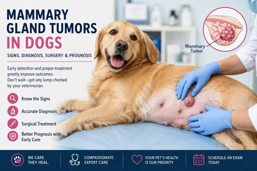
Finding a mammary tumor in dogs can be alarming for any pet owner. Whether you’ve noticed a small lump near your dog’s nipple or a larger swelling along the mammary chain, it’s important not to ignore it. A dog mammary tumor is one of the most common tumors diagnosed in female dogs, particularly those that have not been spayed or were spayed later in life. While not every mammary gland tumor in a dog is cancerous, every new lump should be examined by a veterinarian as early as possible. Early diagnosis helps determine whether the growth is benign or malignant and allows treatment to begin before the disease has a chance to progress.
Because mammary tumors vary widely in their behavior, appearance, and prognosis, it is not possible to determine whether a lump is harmless simply by looking at it or feeling it. A thorough physical examination, diagnostic imaging, and histopathology are often needed to identify the type of tumor and recommend the most appropriate treatment. For many dogs with localized disease, mammary tumor surgery remains the primary treatment option and offers the best opportunity for successful management.
In this guide, you’ll learn what causes mammary tumors in dogs, the signs and symptoms to watch for, how veterinarians diagnose and stage these tumors, treatment options including mammary tumor surgery for dogs, recovery expectations, prognosis, and when to seek veterinary care. If you’re looking for experienced veterinary care, or mammary tumor surgery in Brampton, this article explains what to expect and why early evaluation can make a meaningful difference to your dog’s health.
What Is a Mammary Gland Tumor in a Dog?
Finding a lump along your dog’s abdomen can be worrying, particularly in an older female dog. One possibility is a mammary gland tumor—an abnormal growth arising from tissue within one of the mammary glands.
Mammary tumors are especially common in sexually intact female dogs and are the most common type of neoplasia in this population. They occur much less commonly in spayed females and are rare in male dogs.
The reassuring part is that a mammary tumor does not automatically mean cancer.
Canine mammary tumors represent a diverse group of growths. Approximately half are benign and half malignant, although the exact proportion varies among populations and according to how tumors are classified.
The problem is that we usually cannot determine with certainty whether a mammary lump is benign or malignant simply by looking at it or feeling it.
That is why a new mammary lump deserves veterinary evaluation rather than simply waiting to see whether it grows.
Where Are the Mammary Glands in Dogs?
Most female dogs have four to five pairs of mammary glands, extending in two chains from the chest toward the groin.
A tumor may occur in just one gland, but dogs frequently develop multiple mammary masses. Different masses in the same dog can even represent different tumor types. The glands toward the rear of the abdomen are affected more commonly.
This is why an examination should involve palpation of the entire mammary chain, not just the lump that the owner originally noticed.
Each mass should ideally be identified, measured and recorded.
What Does a Mammary Tumor Look or Feel Like?
Mammary tumors can look very different from one dog to another.
- A small pea-sized lump near a nipple
- One or several firm nodules
- A mass underneath or beside a mammary gland
- A freely movable lump
- A mass attached to deeper tissues
- Rapid enlargement of an existing lump
- Redness or swelling
- Ulceration or bleeding from the surface
- Discharge from a mammary gland
Some tumors remain quite small for a long time. Others can grow rapidly.
Most dogs with localized mammary tumors are otherwise completely well when diagnosed.
The absence of pain does not mean that a lump is harmless.
Many mammary tumors cause little or no discomfort when they are small.
Are Mammary Tumors in Dogs Cancerous?
This is usually the first question an owner asks.
Approximately 50% of canine mammary tumors are benign and 50% are malignant. Merck’s current professional guidance describes mammary tumors as a highly diverse histopathological group rather than one single disease.
Benign tumors generally remain localized and can often be cured by complete surgical removal.
Malignant mammary tumors behave differently. Depending on their type and grade, they may invade surrounding tissues or spread elsewhere in the body.
Common sites of metastasis include:
- Regional lymph nodes
- Lungs
- Liver
- Kidneys
However, even the word malignant does not provide the whole prognosis.
Some mammary carcinomas behave relatively slowly and can have excellent outcomes following complete surgery. Others are considerably more aggressive.
The pathology report therefore becomes extremely important.
Why Early Detection of Mammary Tumors Matters
Early diagnosis gives veterinarians the best opportunity to identify whether a mammary tumor is benign or malignant before it progresses. Smaller tumors are often easier to remove and may offer a better prognosis, making prompt evaluation an important part of your dog’s care.
Why Tumor Size Affects Treatment and Prognosis
This is perhaps the most important practical message for dog owners.
Earlier evaluation is generally better than waiting for the tumor to grow.
Tumor size is an important prognostic factor in canine mammary cancer, and the likelihood of malignancy increases as tumor size increases.
| Tumor size | TNM category |
| Less than 3 cm | T1 |
| 3–5 cm | T2 |
| Greater than 5 cm | T3 |
Lymph-node metastasis or distant metastasis advances the stage regardless of primary tumor size.
From a surgical perspective, smaller tumors can also be easier to remove with appropriate margins and less disruption of surrounding tissue.
So if you find something approximately the size of a pea or grape, there is generally little advantage in deliberately waiting until it becomes much larger before having it assessed.
How Veterinarians Diagnose Mammary Tumors in Dogs
The first step is a thorough physical examination.
We assess not only the obvious mass but the complete mammary chains and regional lymph nodes.
Important characteristics include:
- Number of masses
- Exact location
- Size
- Mobility
- Consistency
- Rate of growth
- Skin involvement
- Ulceration
- Relationship to surrounding tissues
- Regional lymph-node enlargement
The dog’s age, reproductive status, general health and medical history are also important.
Can a Fine-Needle Aspirate Tell Us Whether the Tumor Is Cancerous?
Sometimes—but there are important limitations.
Fine-needle aspiration (FNA) can help determine whether a lump may actually represent another type of skin or subcutaneous tumor. It can also be useful for evaluating suspicious lymph nodes.
However, canine mammary tumors can contain multiple different tissue components. A small needle sample may therefore not reliably distinguish a benign mammary tumor from a malignant one or represent the entire mass.
For most mammary tumors, the definitive diagnosis comes from:
Surgical removal + histopathology.
A veterinary pathologist examines the tissue microscopically and can determine exactly what type of tumor is present.
Why Is Histopathology So Important?
Removing a mammary mass without submitting it for pathology leaves one of the most important questions unanswered:
What exactly did we remove?
Histopathology can provide information about:
- Whether the tumor is benign or malignant
- Histological tumor type
- Tumor grade
- Surgical margins
- Vascular or lymphatic invasion
- Features associated with recurrence or metastasis
Current oncology recommendations emphasize that tumor type, grade and stage are fundamental to treatment planning and prognosis.
For this reason, we recommend histopathology of removed mammary tumors.
Does My Dog Need X-Rays Before Mammary Tumor Surgery?
For a dog with a suspected mammary malignancy, staging is important because finding metastatic disease can change both treatment recommendations and prognosis.
Current veterinary guidance recommends three-view chest radiographs as part of staging canine mammary tumors to evaluate the lungs for visible metastatic disease.
Depending upon the individual dog, additional evaluation may include:
- CBC and serum chemistry
- Urinalysis
- Fine-needle aspiration of suspicious regional lymph nodes
- Abdominal ultrasound when clinically indicated
- Other advanced imaging in selected cases
Not every dog requires every possible diagnostic test.
The staging plan should be appropriate for the individual patient.
What If the Chest X-Rays Are Normal?
Normal thoracic radiographs are reassuring because there is no radiographically detectable pulmonary metastasis.
However, owners should understand an important limitation:
Normal X-rays cannot guarantee that microscopic cancer cells are absent.
Imaging detects disease once lesions become large enough to visualize.
This is one reason the final pathology result remains so important even when preoperative staging looks good.
Mammary Tumor Surgery in Dogs
For most dogs with localized mammary tumors, surgery is the primary treatment.
Surgery serves two purposes:
- Local treatment — removing the tumor.
- Diagnosis — providing tissue for histopathology.
Current Merck and AAHA guidance recognizes several surgical approaches depending upon the individual tumor.
Lumpectomy
Removal of a small tumor with appropriate surrounding tissue.
Simple mastectomy
Removal of the affected mammary gland.
Regional mastectomy
Removal of the affected gland together with anatomically related glands.
Unilateral chain mastectomy
Removal of an entire mammary chain in selected dogs with multiple tumors or disease distribution that warrants a more extensive approach.
The correct surgery depends upon tumor size, location, number, margins and lymphatic anatomy.
Does the Entire Mammary Chain Always Need to Be Removed?
No.
This is an important difference between canine and feline mammary surgery.
Dogs do not automatically benefit from removal of every normal mammary gland simply because one tumor is present. Current evidence does not demonstrate a benefit from prophylactically removing normal unaffected glands in every dog.
A dog with one small, well-localized tumor may require considerably less surgery than a dog with multiple tumors involving several adjacent glands.
The surgical plan should therefore be individualized.
What About the Lymph Nodes?
Mammary glands drain through specific lymphatic pathways, and malignant cells can spread to regional lymph nodes.
The principal nodes of concern include the:
- Axillary lymph nodes
- Superficial inguinal lymph nodes
Suspicious or enlarged lymph nodes may be sampled before surgery.
In some surgical procedures—particularly involving the caudal mammary glands—the superficial inguinal lymph node may be removed along with the associated mammary tissue.
Lymph-node involvement is clinically important because it changes the tumor stage and prognosis.
What Is Inflammatory Mammary Carcinoma?
Inflammatory mammary carcinoma deserves special attention because it behaves very differently from a typical discrete mammary mass.
Instead of one obvious lump, the mammary region may become:
- Diffusely swollen
- Red
- Warm
- Painful
- Thickened
- Inflamed
It can sometimes resemble severe mastitis.
Inflammatory mammary carcinoma is an aggressive form of mammary cancer involving extensive invasion of dermal lymphatics, and conventional surgical removal is generally not beneficial.
A dog with rapidly developing painful, diffuse mammary swelling therefore requires prompt assessment.
Should My Dog Be Spayed When the Mammary Tumor Is Removed?
This question requires more nuance than the traditional answer.
We know that ovarian hormones—particularly estrogen and progesterone—play an important role in canine mammary tumor development.
Early spaying substantially reduces the risk of developing mammary tumors. However, current evidence also recognizes potential health trade-offs associated with early spaying in some breeds, particularly certain large dogs. Therefore, modern recommendations about the age of spaying should be individualized.
But what about an older intact female who already has a mammary tumor?
Historically, the benefit of spaying at the time of mammary tumor removal was debated.
More recent prospective evidence suggests that ovariohysterectomy performed with mammary tumor surgery may reduce the development of new mammary tumors in some dogs, and the 2026 AAHA oncology guidance notes that concurrent or relatively recent ovariohysterectomy may improve survival in a subset of dogs.
Spaying also eliminates the future risk of pyometra.
Therefore, whether to combine spaying with mammary surgery should be decided individually based on:
- Age
- Reproductive status
- Breed
- Number and type of mammary masses
- General health
- Anesthetic considerations
- Expected surgical duration
Also Read: Spay and Neuter in Dogs & Cats: Age Guide for Pet Owners
Is an Older Dog Too Old for Mammary Tumor Surgery?
Age alone is not a disease and is not, by itself, a reason to rule out surgery.
Many mammary tumors occur in older dogs—the average age at diagnosis is approximately 7–11 years.
The more useful question is:
How healthy is this individual dog, and what are the risks and benefits of surgery?
Before anesthesia, we evaluate factors such as:
- Physical examination findings
- Heart and lung status
- Bloodwork
- Kidney and liver function
- Concurrent medical conditions
- Tumor size and extent
- Evidence of metastasis
Additional cardiac or other diagnostic evaluation may be appropriate when abnormalities are detected.
A healthy senior dog with localized disease can be a very different anesthetic candidate from a younger dog with significant systemic illness.
My Dog Has a Heart Murmur. Does That Mean She Cannot Have Surgery?
Not necessarily.
A heart murmur is a physical examination finding, not by itself a complete diagnosis of cardiac function or anesthetic risk.
Its significance depends on the underlying heart disease and its severity.
Depending upon the dog’s history and examination, further evaluation might include chest radiographs, blood pressure assessment, ECG and/or echocardiography before deciding whether and how to proceed with anesthesia.
The decision should be based on the individual patient’s cardiovascular status and the expected benefit of surgery, rather than automatically denying surgery because a murmur is present.
What Is Recovery Like After Mammary Tumor Surgery?
Recovery depends substantially on how extensive the surgery was.
A small local excision is very different from removal of several glands or an entire mammary chain.
After surgery, dogs generally require:
- Appropriate pain medication
- Restricted activity
- An Elizabethan collar or surgical protection to prevent licking
- Monitoring of the incision
- Follow-up examination
- Suture or staple removal when applicable
Activity is commonly restricted for approximately 10–14 days or around two weeks, depending on healing and surgical extent.
Potential complications include swelling, bruising, infection, fluid accumulation and wound dehiscence, particularly following more extensive procedures.
What Is the Prognosis After Mammary Tumor Surgery?
There is no single prognosis for a dog with a mammary tumor.
For benign tumors, complete surgical removal can be curative.
For malignant tumors, prognosis depends on several factors:
- Tumor size
- Clinical stage
- Histologic type
- Tumor grade
- Surgical margins
- Lymph-node involvement
- Vascular invasion
- Presence of distant metastasis
Current veterinary oncology guidance emphasizes that mammary cancers span a very wide biological spectrum—from tumors with excellent long-term outcomes following surgery to highly aggressive cancers.
This is why two dogs who both receive a pathology result containing the word “carcinoma” may nevertheless have very different prognoses.
Does My Dog Need Chemotherapy After Surgery?
Not necessarily.
Dogs with benign tumors do not require chemotherapy.
Many dogs with low-risk malignant tumors that have been completely removed also may not require systemic therapy.
For higher-risk cancers—based on tumor grade, stage, vascular invasion, metastatic disease or aggressive histologic subtype—consultation with a veterinary oncologist may be appropriate.
Evidence for chemotherapy in canine mammary carcinoma is not as definitive as it is for some other canine cancers, so treatment decisions should be individualized rather than assuming every malignant tumor requires chemotherapy.
Can Mammary Tumors Come Back?
Yes.
There are two different possibilities owners should understand:
Local recurrence: tumor returns near the previous surgical site.
Development of a new mammary tumor: another gland develops an independent tumor later.
Because dogs commonly develop multiple mammary tumors—and different tumors in the same dog may even have different pathology—continued examination of the remaining mammary tissue is important after surgery.
Dogs with malignant tumors may also require periodic monitoring of regional lymph nodes and imaging depending upon their pathology results and stage.
Can Mammary Tumors Be Prevented?
Hormonal exposure is strongly associated with mammary tumor development.
Spaying before the first heat substantially reduces mammary tumor risk. However, the ideal age for spaying is no longer considered identical for every dog because reproductive, orthopedic and other cancer risks can vary by breed and adult size.
Therefore, prevention should involve an individualized discussion about the appropriate age for spaying, particularly in large and giant breeds.
For intact females or dogs in which spaying has intentionally been delayed, regular examination of the mammary chains becomes especially important.
Also Read: Spay and Neuter: Health Benefits Beyond Population Control
How to Check Your Dog’s Mammary Glands at Home
Early detection often starts with the owner.
When your dog is relaxed, gently run your fingertips along both mammary chains from the chest toward the groin.
Feel around each nipple for:
- New lumps
- Firm nodules
- Thickening
- Swelling
- Changes in an existing mass
If you find something, don’t repeatedly squeeze or manipulate it.
Instead, arrange a veterinary examination and keep track of when you first noticed it.
A small painless lump is still worth examining.
When Should I See a Veterinarian?
Arrange an examination if you notice any new mammary lump, particularly in an intact or previously late-spayed female dog.
Seek more prompt attention if the mass:
- Is growing rapidly
- Is ulcerated or bleeding
- Has become painful
- Is firmly attached
- Is associated with enlarged lymph nodes
- Is accompanied by weight loss or reduced appetite
- Causes diffuse redness, heat or swelling of the mammary region
Waiting until a tumor becomes large rarely creates an advantage.
Mammary Tumor Evaluation and Surgery in Brampton
At McQueen Animal Hospital in Brampton, we evaluate mammary gland tumors in dogs and develop an individualized plan based on the patient’s health and the characteristics of the tumor.
Depending on the individual dog, this may include:
- Complete examination of both mammary chains
- Pre-anesthetic bloodwork
- Chest X-rays for staging
- Assessment of regional lymph nodes
- Surgical removal of the mammary tumor
- Mammary gland or regional mammary surgery when appropriate
- Histopathology of removed tissue
- Postoperative pain management and monitoring
- Referral or oncology consultation for higher-risk cancers when indicated
Our goal is not simply to remove a lump. It is to determine the appropriate extent of surgery, obtain a diagnosis, assess the risk of spread and help the owner understand what the pathology means for their dog’s future care.
If you’ve discovered a mammary lump on your dog, having it evaluated while it is still small can provide more options than waiting for it to enlarge.
Call McQueen Animal Hospital at 905-455-7387 to schedule an examination.
Frequently Asked Questions About Mammary Tumors in Dogs
Q1. Are mammary tumors in dogs always cancer?
No. Approximately half of canine mammary tumors are benign and approximately half are malignant. The definitive diagnosis usually requires histopathology.
Q2. Should a small mammary lump be removed?
Not every lump automatically requires the same treatment, but a new mammary mass should be evaluated promptly. Tumor size is an important prognostic factor, and earlier surgery can be advantageous when removal is indicated.
Q3. Can a veterinarian tell whether a mammary tumor is cancerous by touching it?
No. Certain characteristics can raise suspicion, but physical examination alone cannot reliably determine whether a mammary tumor is benign or malignant.
Q4. Will a needle aspirate tell us if it is malignant?
Sometimes it provides useful information, but cytology has limitations with mammary tumors. Histopathology of the excised tumor remains the cornerstone of definitive diagnosis.
Q5. Does my dog need chest X-rays before surgery?
For suspected mammary malignancy, thoracic radiographs are recommended for staging because the lungs are an important potential site of metastasis. The exact workup should be individualized.
Q6. Does the entire mammary chain have to be removed?
No. Surgical extent depends on the number, size and location of tumors. There is no proven benefit to routinely removing normal unaffected mammary glands in every dog.
Q7. Is a 12-, 13- or 14-year-old dog too old for mammary tumor surgery?
Not necessarily. Chronological age alone does not determine anesthetic suitability. Overall health, cardiovascular status, organ function, tumor stage and the expected benefit of surgery are more meaningful considerations.
Q8. Can a dog with a heart murmur undergo mammary tumor surgery?
Potentially, yes. A murmur does not automatically prohibit anesthesia. Its cause and severity should be evaluated, and additional cardiac testing may be appropriate before making an anesthetic decision.
Q9. Should an intact dog be spayed at the same time?
It may be appropriate in some dogs. Current evidence suggests concurrent spaying may reduce new mammary tumor development and potentially benefit a subset of dogs, while also preventing pyometra. The decision should be individualized.
Q10. What is the most important thing an owner should do after finding a mammary lump?
Have it examined rather than waiting for it to become large. Earlier diagnosis gives the veterinarian more information and, when surgery is appropriate, often provides better treatment options.
Medically Reviewed by Dr. Harinder Babbar – McQueen Animal Hospital