
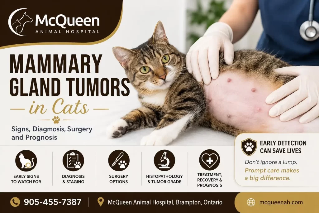
Mammary gland tumors are among the most common cancers in middle-aged and older female cats, and approximately 80 to 90 percent of feline mammary tumors are malignant. Unlike in dogs, where many mammary tumors are benign, feline mammary tumors behave aggressively and can spread to regional lymph nodes, the lungs and other organs if left untreated. Tumor size at diagnosis is one of the strongest predictors of outcome, which is why any new lump along a cat’s mammary chain deserves prompt veterinary evaluation rather than a “wait and see” approach. This guide covers how mammary tumors are diagnosed and staged, what surgery involves, what recovery looks like, and what to expect from evaluation and treatment at McQueen Animal Hospital in Brampton.
What Is a Mammary Gland Tumor in a Cat?
Discovering a lump along your cat’s abdomen or near a nipple can be alarming. Unlike dogs, in whom a significant proportion of mammary tumors are benign, approximately 80–90% of mammary tumors in cats are malignant. Most malignant feline mammary tumors are carcinomas, and they may invade surrounding tissue or spread to regional lymph nodes, the lungs and other organs.
A mammary gland tumor develops from abnormal cells within the milk-producing tissue of one or more mammary glands. These tumors may begin as very small, firm nodules but can enlarge, ulcerate and metastasize if they are not addressed.
Mammary tumors most commonly affect middle-aged and older female cats, particularly those that remain intact or were spayed later in life. They are uncommon in male cats, but a mammary lump in a male cat still requires prompt examination.
The most important message for cat owners is:
A mammary lump in a cat should not be ignored simply because it is small, painless or not affecting the cat’s behaviour.
Early evaluation provides the greatest opportunity to stage the disease properly and, when surgery is appropriate, to remove the tumor while it is still relatively small.
Why Are Mammary Tumors More Serious in Cats Than in Dogs?
Mammary tumors behave differently in cats and dogs.
In cats, most mammary tumors are malignant, and many behave aggressively. They may grow into nearby skin or underlying tissue and spread through lymphatic vessels to regional lymph nodes. The lungs are another important site of metastasis.
This biological behaviour has several practical consequences:
- A feline mammary lump deserves prompt investigation.
- Prolonged “wait and see” monitoring is generally not advisable.
- Staging before surgery is important.
- Removing only the visible lump may not provide adequate local control.
- Histopathology is essential after surgery.
- Follow-up monitoring remains important even after successful removal.
A small tumor does not guarantee that the growth is benign. However, cats treated while tumors are still small generally have more favourable outcomes than cats presented with large or advanced masses.
Where Are the Mammary Glands in Cats?
Cats usually have four pairs of mammary glands, arranged in two chains extending from the chest toward the groin.
A tumor may affect:
- One mammary gland
- Several glands on the same side
- Both mammary chains
- The tissue underneath or adjacent to a nipple
The rear, or caudal, mammary glands are commonly involved, although tumors can arise anywhere along either chain.
Because a cat may have more than one tumor, the veterinarian should examine and palpate both complete mammary chains, not only the lump first noticed by the owner.
Each mass should ideally be:
- Located
- Measured
- Assessed for mobility
- Checked for skin involvement
- Recorded in the medical record
Regional lymph nodes should also be evaluated.
What Causes Mammary Tumors in Cats?
The exact cause of feline mammary cancer is not completely understood, but reproductive hormones play an important role.
Intact female cats have a substantially higher risk of mammary carcinoma than cats spayed early in life. One influential study found that cats spayed before six months of age had approximately a 91% reduction in risk, while those spayed before one year had approximately an 86% reduction, compared with intact females.
The protective effect declines as exposure to ovarian hormones increases. This is one of the strongest medical reasons for recommending spaying before a cat reaches sexual maturity.
Hormonal medications containing progestogens may also contribute to abnormal mammary tissue growth in cats. A separate benign condition called mammary fibroadenomatous hyperplasia may occur in young intact cats or cats exposed to progestogens and can occasionally resemble neoplasia. A veterinary examination is therefore necessary rather than assuming every mammary enlargement is cancer.
What Does a Mammary Tumor Look or Feel Like?
Feline mammary tumors can have different appearances.
An owner may notice:
- A small, pea-sized lump near a nipple
- A firm nodule under the skin
- Thickening beneath one mammary gland
- Several small nodules along the mammary chain
- A poorly movable or attached mass
- A rapidly enlarging growth
- Redness or swelling
- Ulceration
- Bleeding
- Discharge from the affected area
Mammary tumors are often firm and may initially be painless. A cat can continue eating, grooming, playing and behaving normally even while carrying a malignant tumor.
The absence of pain or illness does not mean that the mass is harmless.
As the tumor progresses, the skin may break down and become infected or uncomfortable. Cats with metastatic or advanced disease may develop weight loss, reduced appetite, lethargy or difficulty breathing.
Common Signs of Mammary Cancer in Cats
Many cats initially show no sign other than a small lump.
Possible signs include:
- One or more mammary nodules
- Gradual or rapid tumor growth
- Firm attachment to surrounding tissue
- Red or inflamed skin
- Ulceration or bleeding
- Discharge from a mammary gland
- Pain or sensitivity
- Reduced appetite
- Weight loss
- Reduced activity
- Enlarged regional lymph nodes
- Coughing or difficulty breathing if disease has spread to the lungs
A breathing abnormality in a cat with a known mammary tumor requires prompt assessment.
Why Tumor Size Is One of the Most Important Prognostic Factors
Tumor size at diagnosis is one of the most consistently recognized prognostic factors in feline mammary carcinoma.
Cats with tumors measuring less than 2 cm generally have considerably better outcomes than cats with larger tumors. Merck’s current veterinary guidance reports median survival exceeding three years for some cats with tumors smaller than 2 cm, compared with markedly shorter survival for cats with tumors larger than 3 cm. Other published studies have reported some variation in exact survival times, but the overall relationship is consistent: larger tumors are associated with a worse prognosis.
There are several reasons why earlier treatment may help:
- Smaller tumors are generally easier to remove with appropriate margins.
- They are less likely to have invaded extensive surrounding tissue.
- They may have had less time to spread through lymphatic or blood vessels.
- Surgery is often less extensive than it would be after substantial growth.
- The cat may be in better overall condition at the time of treatment.
This does not mean that every small tumor will behave favourably or that every large tumor has already metastasized. Tumor grade, lymphovascular invasion, lymph-node status, surgical margins and other factors also matter.
However, the evidence strongly supports one practical recommendation:
Do not deliberately wait for a feline mammary lump to become larger before having it examined.
Feline Mammary Tumor Size and Prognostic Significance
| Largest tumor diameter | General interpretation |
|---|---|
| Less than 2 cm | Generally associated with the most favourable outcomes |
| 2–3 cm | Intermediate risk; prognosis depends heavily on stage, grade and other findings |
| Greater than 3 cm | Associated with a substantially more guarded prognosis |
These ranges should not be used as a substitute for staging and histopathology. They help explain why early examination is important, but they do not predict the outcome of every individual cat.
How Are Mammary Tumors in Cats Diagnosed?
Diagnosis begins with a thorough pet consultation and examination.
The veterinarian evaluates:
- The number of masses
- The location of each mass
- The size of the largest tumor
- Whether the tumor is freely movable or attached
- Whether the skin is involved
- Whether ulceration is present
- The condition of both mammary chains
- Regional lymph-node enlargement
- The cat’s general health
The cat’s age, reproductive history, previous hormone exposure, rate of tumor growth and any changes in appetite, weight or breathing are also relevant.
The physical examination can establish the level of suspicion, but it cannot reliably determine the exact tumor type or grade.
Can a Needle Aspirate Diagnose a Mammary Tumor?
A fine-needle aspirate may be useful in some cases. It can help distinguish a mammary tumor from another type of skin or subcutaneous mass, and it may be helpful for evaluating suspicious lymph nodes.
However, feline mammary tumors may contain several areas with different microscopic characteristics. A small needle sample may not represent the entire tumor and may not provide enough information to determine tumor grade or surgical margins.
For most suspected feline mammary tumors, the definitive diagnosis comes from:
Surgical removal followed by histopathology.
The removed tissue is examined microscopically by a veterinary pathologist.
Types of Mammary Tumors in Cats
Not all feline mammary tumors behave in exactly the same way.
Most malignant feline mammary tumors are carcinomas or adenocarcinomas. Pathologists may describe different growth patterns or subtypes, including:
- Tubular carcinoma
- Papillary carcinoma
- Solid carcinoma
- Cribriform carcinoma
- Other mixed or less common patterns
Some tumors are well differentiated, meaning their cells retain more recognizable mammary features. Others are poorly differentiated and may behave more aggressively.
Pet owners do not need to interpret every microscopic term alone. The more useful questions are:
- Is the tumor benign or malignant?
- What type of malignant tumor is present?
- What is the tumor grade?
- Were the margins complete?
- Is there lymphatic or vascular invasion?
- Were lymph nodes involved?
- Does the pathology suggest a high risk of recurrence or metastasis?
The scientific name of the tumor is important, but the pathology report’s overall assessment is what guides treatment and follow-up.
Why Histopathology Is Essential After Mammary Tumor Surgery
Surgery removes the visible disease, but histopathology tells us what was removed.
A pathology report may provide information about:
- Benign versus malignant disease
- Histologic tumor type
- Tumor grade
- Mitotic activity
- Degree of cell differentiation
- Lymphatic or vascular invasion
- Surgical margins
- Features associated with recurrence or metastasis
These findings help the veterinarian determine whether:
- Surgery may be sufficient
- Closer monitoring is required
- Additional staging should be performed
- Referral to a veterinary oncologist should be considered
- Chemotherapy should be discussed
Every removed feline mammary tumor should ordinarily be submitted for veterinary pathology and biopsy testing. Without pathology, we may remove the visible mass but remain uncertain about its biological behaviour and the cat’s future risk.
What Do “Tumor Grade,” “Margins” and “Lymphatic Invasion” Mean?
Tumor Grade
Tumor grade describes how abnormal and aggressive the cancer appears under the microscope.
A higher-grade tumor generally has:
- More abnormal-looking cells
- Faster cell division
- Less organized tissue
- A greater tendency to recur or metastasize
Grade is only one part of prognosis and should be interpreted alongside tumor size, stage and surgical margins.
Surgical Margins
The pathologist examines the edges of the removed tissue to assess whether cancer cells extend to or near the surgical boundary.
Clean or complete margins mean that no tumor cells were identified at the cut edge. This is favourable, although it cannot guarantee that microscopic disease does not exist elsewhere.
Incomplete or involved margins mean tumor cells reach the edge of the submitted tissue. Depending on the case, further surgery, oncology consultation or close monitoring may be considered.
Lymphatic or Vascular Invasion
This means cancer cells were identified inside lymphatic channels or blood vessels.
Lymphovascular invasion can indicate a greater potential for metastatic spread and may influence prognosis and decisions about additional treatment.
Staging Before Mammary Tumor Surgery
Because feline mammary carcinomas have a meaningful risk of metastasis, staging is an important part of treatment planning.
Depending on the cat’s condition and tumor characteristics, staging may include:
- Complete blood count
- Serum chemistry profile
- Urinalysis
- Three-view chest radiographs
- Evaluation of regional lymph nodes
- Fine-needle aspiration or biopsy of suspicious lymph nodes
- Abdominal ultrasound in selected patients
- Additional imaging where clinically indicated
Bloodwork, run through our on-site veterinary laboratory testing services, does not diagnose mammary cancer, but it helps assess the cat’s overall health and anesthetic suitability. In selected patients, our advanced veterinary ultrasound imaging services are also used to evaluate the abdomen for signs of internal spread.
Chest radiographs, captured through our digital pet radiography diagnostic imaging, are used to look for visible pulmonary metastases. Regional lymph nodes are assessed because mammary cancer often spreads through lymphatic pathways.
Why Chest X-Rays Can Be Normal Even When Cancer Has Spread
Owners are understandably relieved when preoperative chest X-rays show no evidence of metastasis.
A normal radiographic study is encouraging because no radiographically detectable lung metastases were found. However, chest X-rays have an important limitation:
They cannot identify individual cancer cells or microscopic metastatic deposits that are too small to visualize.
Imaging detects lesions only once they reach a sufficient size or density.
A normal chest X-ray should therefore be interpreted as:
No visible pulmonary metastasis was detected at the time of imaging.
It should not be interpreted as an absolute guarantee that no microscopic cancer cells exist anywhere in the body.
This is why surgery, histopathology, lymph-node assessment and follow-up monitoring all remain important even when preoperative imaging is normal.
Surgery for Mammary Tumors in Cats
Our comprehensive pet surgery services in Brampton remain the primary local treatment for most cats with operable mammary tumors and no contraindication to anesthesia.
Unlike dogs, cats generally benefit from a more aggressive surgical approach because feline mammary tumors have a high malignant rate and may extend microscopically beyond the visible lump. ACVS and veterinary oncology literature describe radical or chain mastectomy as an important component of local treatment in cats.
Depending on the distribution of disease, surgery may include:
Unilateral Chain Mastectomy
Removal of the entire mammary chain on the affected side.
Bilateral Staged Chain Mastectomy
Removal of one mammary chain, followed by removal of the opposite chain after the first surgery has healed.
Bilateral surgery is commonly staged rather than removing both chains in one procedure because staged operations may reduce wound tension and make postoperative recovery more manageable.
The appropriate surgical plan depends on:
- Number of tumors
- Tumor location
- Whether one or both chains are involved
- Previous mammary surgery
- Tumor attachment and skin involvement
- Regional lymph-node findings
- Staging results
- Overall health of the cat
Why We Recommend Removing the Entire Mammary Chain in Many Cats
This is one of the most common questions cat owners ask.
Removing only the visible lump may appear less invasive. However, feline mammary cancer may extend microscopically beyond what can be seen or felt. Additional small tumors or microscopic cancer cells may be present elsewhere in the affected chain.
More extensive mammary surgery has been associated with better local control and a lower rate of recurrence than conservative removal of only the visible mass.
The objective is not merely to remove the lump that is visible today. It is to:
- Remove the primary tumor
- Obtain appropriate margins
- Reduce the amount of potentially affected mammary tissue left behind
- Lower the risk of local recurrence
- Provide tissue for complete histopathological evaluation
That does not mean every cat automatically receives exactly the same operation.
A surgical plan must account for:
- The location and number of tumors
- Whether disease is unilateral or bilateral
- Tumor size
- Skin involvement
- Previous surgeries
- The cat’s general health
- The practical balance between oncological benefit and surgical risk
For many cats, an entire affected mammary chain is removed. For cats with disease affecting both sides, removal of both chains may be considered as two staged procedures.
Why Not Remove Only the Lump?
A simple lumpectomy removes the palpable mass but leaves the rest of the affected mammary chain intact.
This may be insufficient because:
- Microscopic tumor extension may remain.
- Other very small tumors may not yet be palpable.
- Local recurrence may be more likely.
- Adequate margins can be difficult to achieve around a limited excision.
The least invasive-looking operation is not always the operation that provides the best cancer control.
The final recommendation should nevertheless be individualized after examination and staging.
What About the Regional Lymph Nodes?
Feline mammary cancer may spread through lymphatic vessels to regional lymph nodes.
Depending on the tumor’s location, lymphatic drainage may involve:
- Axillary lymph nodes
- Inguinal lymph nodes
- Other connected regional nodes
Suspicious lymph nodes may be:
- Palpated during examination
- Evaluated by fine-needle aspiration
- Removed at surgery when appropriate
- Submitted separately for histopathology
Lymph-node involvement changes the clinical stage and generally carries a less favourable prognosis. A recent review summarized studies in which cats with nodal metastasis had shorter median survival than those without known nodal involvement.
Does My Cat Need Chemotherapy for Mammary Cancer?
Not every cat with a mammary tumor automatically requires chemotherapy.
Chemotherapy may be discussed when pathology or staging identifies higher-risk disease, including:
- High tumor grade
- Lymphatic or vascular invasion
- Lymph-node metastasis
- Incomplete surgical margins
- Large primary tumor
- Multiple aggressive tumors
- Documented distant metastasis
- Rapid recurrence
The evidence supporting chemotherapy for feline mammary carcinoma is less definitive than for some other cancers. Decisions should therefore be individualized after reviewing the pathology report and clinical stage.
A veterinary oncology consultation may help owners understand:
- Whether chemotherapy is reasonable
- What potential benefit is expected
- The treatment schedule
- Possible adverse effects
- Monitoring requirements
- Alternatives, including surveillance or palliative care
Veterinary chemotherapy is generally designed to preserve quality of life rather than deliver the maximum dose tolerated in human oncology.
What to Expect If Chemotherapy Is Recommended
The exact protocol depends on the oncologist, tumor characteristics and the cat’s health.
Treatment may involve:
- Periodic hospital visits
- Injectable or oral medication
- Blood testing before treatments
- Monitoring appetite, activity and gastrointestinal signs
- Dose adjustment when necessary
Many cats tolerate veterinary chemotherapy reasonably well, although possible adverse effects include:
- Reduced appetite
- Vomiting
- Diarrhea
- Lethargy
- Temporary reduction in white blood cells
The veterinarian or oncologist should discuss anticipated benefit, risks and goals before treatment begins.
Recovery After Mammary Chain Surgery
Recovery depends on whether one gland, one chain or both chains in staged procedures are removed.
Mammary chain surgery creates a relatively long incision, and careful postoperative management is important.
After surgery, a cat may require:
- Injectable and oral pain medication
- An Elizabethan collar or properly fitted surgical recovery garment
- Restricted activity
- A quiet indoor recovery area
- Incision monitoring
- Follow-up examinations
- Suture or staple removal when applicable
- Histopathology review when results become available
Potential complications include:
- Swelling
- Bruising
- Fluid accumulation
- Infection
- Incisional separation
- Reduced appetite
- Discomfort
- Skin tension or delayed healing
Contact the hospital promptly if the incision opens, becomes increasingly red or swollen, develops discharge, or if the cat stops eating, becomes markedly lethargic or appears painful.
General Recovery Timeline
First 24 Hours
Your cat may be sleepy after anesthesia. Appetite may be reduced temporarily. Pain control, warmth and observation are important.
Days 2–3
The cat should gradually become brighter and more interested in food. Mild swelling or bruising may occur, but worsening pain, discharge or marked swelling should be reported.
Days 4–7
The incision should remain closed and dry. Activity must still be restricted even if the cat appears energetic.
Days 10–14
Many skin incisions are substantially healed by this stage. A follow-up examination may be performed, and external sutures or staples may be removed when appropriate.
After Two Weeks
Activity may gradually return to normal after veterinary approval. Cats undergoing staged bilateral surgery may then continue recovery before planning the second procedure.
This timeline is a general guide. Recovery varies according to the surgery performed and the individual cat.
What Is the Prognosis for Cats With Mammary Cancer?
There is no single prognosis for every feline mammary tumor.
Important prognostic factors include:
- Tumor size
- Clinical stage
- Histologic tumor type
- Tumor grade
- Surgical margins
- Lymph-node involvement
- Lymphovascular invasion
- Presence of distant metastasis
- Extent of surgery
- Overall health of the cat
Cats with small, localized tumors that are treated aggressively before metastasis generally have the most favourable outlook.
Cats with large tumors, lymph-node metastasis, high-grade disease or visible distant spread have a more guarded prognosis.
The pathology report and staging findings should be interpreted together. One isolated feature rarely tells the entire story.
What Is the Life Expectancy for a Cat With Mammary Cancer?
This is one of the most common and difficult questions owners ask.
Published survival figures vary among studies because cats differ in tumor size, stage, surgery, pathology and follow-up treatment.
Current veterinary references describe a clear overall pattern:
- Cats with tumors under 2 cm can sometimes have median survival measured in years.
- Tumors between 2 and 3 cm carry an intermediate prognosis.
- Tumors larger than 3 cm have often been associated with median survival measured in months, although exact estimates vary between studies.
These are population averages, not predictions for an individual cat.
A particular cat’s prognosis must account for:
- Whether metastasis was found
- Tumor grade
- Lymph-node status
- Completeness of surgery
- Lymphovascular invasion
- Overall health
- Whether additional therapy is undertaken
It is more useful to discuss prognosis after staging and histopathology than to provide an exact life-expectancy number before those results are available.
Can Mammary Tumors in Cats Return?
Yes.
Local Recurrence
Cancer regrows near the original surgical site.
A New Mammary Tumor
Another area of remaining mammary tissue develops a separate tumor.
Regional Metastasis
Disease spreads to nearby lymph nodes or surrounding tissues.
Distant Metastasis
Cancer spreads to organs such as the lungs.
The risk depends on stage, grade, margins, surgical extent and other biological factors.
Follow-up examinations may include:
- Palpation of surgical sites and remaining mammary tissue
- Regional lymph-node assessment
- Chest imaging
- Additional testing based on the pathology report and clinical signs
Can Mammary Tumors in Cats Be Prevented?
No preventive measure eliminates all risk, but the spay and neuter surgery benefits are well documented, and early spaying substantially reduces the likelihood of developing feline mammary carcinoma.
Cats spayed before six months of age have been reported to have about a 91% reduction in risk, while cats spayed before one year have an approximately 86% reduction compared with intact females. These figures are among the reasons early spaying is considered one of the most effective steps toward reducing your pet’s cancer risk over a lifetime.
Other practical steps include:
- Avoiding unnecessary progestogen hormone treatment
- Scheduling regular veterinary examinations
- Checking the mammary chains at home
- Arranging prompt assessment of any new lump
Early detection is not true prevention, but it can significantly influence the treatment options available.
How to Check Your Cat’s Mammary Glands at Home
Once a month, when your cat is relaxed, gently run your fingertips along both sides of the abdomen from the chest toward the groin.
Feel around each nipple for:
- A new lump
- Firm nodules
- Thickening
- Swelling
- Skin changes
- Discharge
- Changes in an existing mass
Do not repeatedly squeeze or manipulate a lump.
Record where you noticed it and arrange a veterinary examination.
A small, painless mass is still worth investigating.
When Should I See a Veterinarian?
Arrange an examination after discovering any new lump along your cat’s mammary chain.
Seek prompt attention if the mass:
- Is growing quickly
- Has become ulcerated
- Is bleeding
- Produces discharge
- Is firmly attached
- Is associated with swelling
- Appears painful
- Is accompanied by weight loss
- Is associated with reduced appetite
- Is accompanied by coughing or difficulty breathing
Waiting until a feline mammary tumor becomes large rarely provides an advantage.
What Happens After Finding a Mammary Lump? A Practical Decision Pathway
Step 1: Arrange a Veterinary Examination
The veterinarian examines both mammary chains, the mass, lymph nodes and the cat’s overall condition.
Step 2: Measure and Document the Tumor
Tumor size and location are recorded because size is an important prognostic factor.
Step 3: Perform Health and Staging Tests
This may include bloodwork, urinalysis, chest radiographs and lymph-node assessment.
Step 4: Discuss Surgical Options
The veterinarian recommends the appropriate extent of mammary surgery based on tumor distribution and patient health.
Step 5: Submit Removed Tissue for Histopathology
The pathology report identifies tumor type, grade, margins and additional prognostic factors.
Step 6: Review the Pathology Report
The veterinarian explains whether further treatment, oncology consultation or monitoring is advisable.
Step 7: Continue Follow-Up Care
The surgical area, remaining tissue, lymph nodes and lungs may require ongoing monitoring.
Mammary Tumor Evaluation and Surgery in Brampton
At McQueen Animal Hospital in Brampton, we evaluate cats with mammary gland tumors and develop individualized diagnostic and surgical plans based on each patient’s findings and general health.
Depending on the individual cat, care may include:
- Complete examination of both mammary chains
- Measurement and documentation of all masses
- Pre-anesthetic bloodwork
- Chest X-rays for staging
- Regional lymph-node assessment
- Surgical planning
- Unilateral mammary chain removal when indicated
- Staged bilateral chain surgery when appropriate
- Histopathology of all removed tissue
- Postoperative pain management
- Incision monitoring and follow-up
- Oncology referral for higher-risk cancers when appropriate
Our objective is not simply to remove a visible lump. It is to determine:
- Whether the tumor may have spread
- What surgical approach offers appropriate local control
- What the pathology means
- Whether additional treatment should be considered
- How the cat should be monitored afterward
If you have discovered a mammary lump on your cat, having it examined while it is still small may provide considerably more options than waiting for it to enlarge.
Call McQueen Animal Hospital at 905-455-7387 to schedule an examination.
Frequently Asked Questions About Mammary Tumors in Cats
Are mammary tumors in cats usually cancerous?
Yes. Approximately 80–90% of feline mammary tumors are malignant, and many behave aggressively. Histopathology is required to establish the exact diagnosis.
Should I wait to see whether the lump grows?
Generally, no. Tumor size is an important prognostic factor, and earlier diagnosis may allow more effective surgery while the tumor is smaller.
Can a veterinarian tell whether the lump is cancer by touching it?
No. Certain physical features may increase concern, but examination alone cannot determine the exact tumor type or grade.
Can a needle aspirate confirm the diagnosis?
It may provide useful preliminary information, but feline mammary tumors can be heterogeneous. Definitive diagnosis usually requires histopathology of surgically removed tissue.
Why does my cat need chest X-rays?
Chest radiographs look for visible metastasis in the lungs. Normal X-rays are reassuring but cannot rule out microscopic disease.
Why might the entire mammary chain need to be removed?
Feline mammary cancer may extend microscopically beyond the visible lump. Removing the affected chain generally offers better local control than removing only the palpable mass.
Does removing the whole chain guarantee the cancer will not return?
No operation can guarantee that cancer will never recur or metastasize. More extensive surgery may improve local control, but outcome also depends on stage, grade, margins, lymph-node status and other factors.
Why are both mammary chains not always removed at once?
Removing both chains simultaneously can create substantial wound tension and may make recovery more difficult. When bilateral removal is needed, the procedures are often staged.
Is an older cat too old for surgery?
Not necessarily. Chronological age alone does not determine anesthetic suitability. Overall health, organ function, cardiac status, disease stage and expected benefit are more meaningful considerations.
What if my cat has kidney disease or a heart murmur?
These conditions do not automatically rule out surgery. Additional evaluation may be needed to determine anesthetic risk and modify the plan appropriately.
How long does recovery take?
The incision commonly requires around 10–14 days for initial skin healing, although recovery depends on the extent of surgery and the individual cat.
Will my cat need chemotherapy?
Not always. Chemotherapy may be considered for higher-risk tumors, lymph-node involvement, metastasis, aggressive pathology or other concerning findings.
What does a clean surgical margin mean?
It means the pathologist did not identify tumor cells at the cut edge of the submitted tissue. This is favourable but does not absolutely guarantee that no microscopic disease exists elsewhere.
What is tumor grade?
Grade describes how aggressive the cancer appears under the microscope. Higher-grade tumors generally carry a greater risk of recurrence or metastasis.
Why is lymph-node involvement important?
Cancer in a regional lymph node indicates metastatic spread and changes the tumor’s clinical stage and prognosis.
Can male cats develop mammary tumors?
Yes, although mammary tumors are much less common in male cats. Any mammary mass in a male cat still requires investigation.
Can spaying prevent mammary cancer?
Early spaying substantially reduces the risk, particularly when performed before six months to one year of age.
Can a mammary mass be something other than cancer?
Yes. Other conditions, including benign tumors, inflammation and hormone-associated mammary hyperplasia, can cause mammary enlargement. However, because most feline mammary tumors are malignant, examination should not be delayed.
Can mammary cancer spread even when my cat seems healthy?
Yes. Cats may appear completely normal despite microscopic or early metastatic disease.
What is the most important thing to do after finding a lump?
Arrange a veterinary examination promptly rather than waiting for the lump to become larger.
Medically reviewed by Dr. Harinder Babbar, DVM
Veterinarian & Medical Director
McQueen Animal Hospital, Brampton, Ontario